 Fundus photography uses a specialized, sophisticated digital camera with powerful lenses that focus on the ocular structures of your inner eye. Your fundus is found at the back of your eyeball, which is where your retina is located. A classic fundus photo will show your optic nerve, main retinal blood vessels and macula. These images are used to record and diagnose many eye conditions, including inspecting closely for the effects of diabetes.
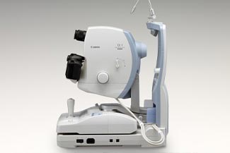
At Hartsdale Family Eyecare, our office is equipped with Canon’s Retinal Camera with advanced fundus photography. Dr. Arlene Schwartz, our professional optometrist, is skilled and experienced in recording and analyzing these pictures as a part of our comprehensive eye exams. We will document the images of your fundus and compare them during follow-up visits to our Hartsdale, New York, office. This is a highly effective way to detect changes in your eye health, especially diabetic eye conditions, as well as pay attention to retinal disease, macular holes, macular degeneration and retinal detachment or tears.
How does fundus photography work?
Dilating eye drops will first be placed in your eyes, as light rays from the fundus camera enter your eye through the pupil. After the drops take effect, we will ask you to sit at our compact Canon camera, resting on the chin rest with your forehead against the bar. Once we align and focus the camera, you’ll see a quick flash of light, which generates the fundus photograph.
What eye conditions can be detected with fundus photography?
A fundus photo shows our Hartsdale eye doctors a comprehensive, detailed view of your retina. By comparing images from visit to visit, any changes in your retinal health will be noticed efficiently. In particular, Dr. Arlene Schwartz uses fundus photography to keep watch on the signs of diabetes.
Recorded fundus pictures are ideal for tracking the progression of diabetic retinopathy, in which uncontrolled diabetes damages the retina, or macular swelling (macular edema). We will also pay attention to the effects of glaucoma on the optic nerve. In comparison to direct eye examinations, fundus photography makes many of the ocular symptoms caused by diabetes easier to visualize.